Article Title: Evaluation of Possible Radioprotective Action of Rosmarinus officinalis L. in Swiss albino Mice
Author Names: Garima Sancheti and P.K. Goyal
Institution & Address: Radiation & Cancer Biology Laboratory
Department of Zoology,
University of Rajasthan,
Jaipur – 302 004 (India)
Key Words: Hematology, Gamma radiation, Glutathione, Lipid peroxidation, Rosmarinus officinalis, Swiss albino mice
First Author: Dr. Garima Sancheti (Ph. D., Alumnus)
Radiation & Cancer Biology Laboratory
Department of Zoology,
University of Rajasthan,
Jaipur – 302 004 (India)
Email: garimasancheti@rediffmail.com
Corresponding Author: Dr. P. K. Goyal
Radiation & Cancer Biology Laboratory
Department of Zoology,
University of Rajasthan,
Jaipur – 302 004 (India)
Tel: 0099-0141-2651199
Email: pkgoyal2002@rediffmail.com
ABSTRACT
Ionizing radiation is one of the most horrible ecological crisis to which living beings are subjected. The inadvertent exposure of human to various sources of radiation causes ionization of molecules that set off potential damaging reactions leading to different health disorders. Thus, it is considered important to explore radio-protective compounds that would be effective and non-toxic at optimum doses. The present study investigates the effect of Rosmarinus officinalis L. against radiation-induced hematological alterations in mice. For this purpose, adult Swiss albino mice were irradiated with 6 Gy gamma rays in the presence (experimental) or absence (control) of rosemary leaves extract (1000 mg/kg body wt.). Animals were autopsied and blood sample was collected at various post-autopsy intervals (i.e., between 24 hours to day 30). Treatment of animals with rosemary extract prior to irradiation was found to reduce the symptoms of radiation sickness in comparison to control group. A decrease in the number of Red Blood Corpuscle (RBC) and White Blood Cell (WBC) counts, hemoglobin content and hematocrit percentage was recorded in the control group; whereas a recovery pattern was scored in experimental animals. A normal value of hematological parameters was regained by the last autopsy interval (i.e., day 30). Irradiation resulted in a significant increase in lipid peroxidation level and a decline in reduced glutathione level in the blood. Conversely, prior treatment of animals with rosemary extract exhibited a significant decrease in lipid peroxidation level and an increase in the glutathione content. The results of our investigation suggest the radioprotective effect of rosemary extract on hematological and biochemical alterations in mice.
KEYWORDS
Hematology, Gamma radiation, Glutathione, Lipid peroxidation, Rosmarinus officinalis L., Swiss albino mice
ABBREVIATIONS
Body weight (b. wt.), Double Distilled Water (DDW), Glutathione (GSH), Hematocrit (Hct), Hemoglobin (Hb), Lipid Peroxidation (LPx), Red Blood Corpuscle (RBC), Rosemary Extract (RE), White Blood Cell (WBC)
INTRODUCTION
The twentieth century has seen an increasing use of nuclear energy in industrial, medical, engineering and scientific research that have raised the problem of radiation hazards to living beings. Thus, the development of effective radioprotectors and radio recovery drugs is of great importance in view of their potential application during both planned (i.e., radiotherapy) and unplanned radiation exposure (i.e., in the nuclear industry, natural background radiation) (Bump and Malaker, 1998).
Extensive work has been carried out in the field of chemical radioprotection during the last few decades but no compound has been found that can provide optimum protection in clinical field without toxicity. Therefore, the search for alternative sources as ideal radioprotective agents, including plants, has been going on (Lam and Ng, 2002; Song et al., 2003).
Plants have been utilized since time immemorial for curing various diseases. Even today, nearly 70% of the world’s population is dependent on plants for handling health disorders (Fabricant and Farnsworth, 2001). Fortunately, many plant derived antioxidant nutrients and phytochemicals have the advantage of low toxicity, therapeutic potential and are protective when administered at pharmacological doses.
Recently, interest has developed in exploring potential drugs of plant origin for the modification of radiation effects. Because of their low toxicity, naturally occurring dietary components offer opportunities for development as radioprotectors (Sarkar, 2004). Plant extract such as that of Panax ginseng (Kim et al., 1993), Spirulina platenis (Qishen et al., 1997), Podophyllum (Goel et al., 1999), Ocimum sanctum (Uma devi et al., 2000), Moringa oleifera (Rao et al., 2001), Mentha arvensis (Jagetia and Baliga, 2002), Adhatoda Vasica (Kumar et al., 2003), Emblica officinalis (Singh et al., 2005) have been found to have an advantage over the synthetic compounds in terms of low/no toxicity at the effective dose with minimum or no side effects. Also, phytochemicals like Caffeine, Genistein and Melatonin have multiple physiological effects as well as antioxidant activity that result in radio-protective role (Zhang et al., 1997; Landauer et al., 2003; Reiter and Tan, 2002).
Rosmarinus officinalis L. is one of the most popular species of the family Lamiaceae, recognized mainly for its culinary and antioxidant properties. It is widely cultivated evergreen and shrubby perennial plant, a native to the Mediterranean region. It has been reported that the aqueous extract of leaves of R. officinalis significantly inhibit the induction of skin papillomas in Swiss albino mice (Sancheti and Goyal, 2006a,b). The antioxidant and pharmacological properties of Rosemary make it an excellent candidate for investigation as a radioprotector. The present study has been undertaken to evaluate the effect of Rosmarinus officinalis leaves extract on radiation induced hematological and biochemical alterations in Swiss albino mice.
MATERIALS AND METHODS
Animals
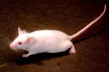
Random bred male Swiss albino mice (Mus musculus), 6-8 weeks old weighing 20-24 g, maintained at the animal house of the Department, were used for the present study (Fig-1). The colony was maintained under controlled conditions of temperature and light (Light: dark, 10 hrs: 14 hrs.). The animals were provided standard mice feed (procured from Hindustan Lever Ltd., India) and water ad libitum.
Animal care and handling were performed according to the guidelines set by the World Health Organization (WHO), Geneva, Switzerland and the INSA (Indian National Science Academy), New Delhi, India. The present study is approved by the Departmental Animal Ethical Committee.
Irradiation
The animals were whole-body exposed to gamma radiation by Cobalt teletherapy unit (Co-60) source (dose rate= 0.85 Gy/min) at a distance of 80 cm, at the Cancer Treatment Centre, Radiotherapy Department, SMS Medical College & Hospital, Jaipur.
Preparation of plant extract
The identification of the plant Rosmarinus officinalis (family: Lamiaceae) (Voucher Specimen No: DDC/2001/DEPTBT/ACHARYA2430) was done by a competent botanist from Department of Botany, Danielson College, Chhindwara, Madhya Pradesh, India. The plant extract was prepared by extracting the non-infected leaf powder of Rosmarinus officinalis with double distilled water by refluxing for 36 hrs at 50-600 C. The extract thus obtained was evaporated in an incubator. For the required dose level (1000 mg/ kg b. wt.), the aqueous extract was dissolved in vehicle, double distilled water; accordingly 0.1 ml of rosemary extract/day was administered to each animal by oral gavage (Fig-2).
Dose selection of Rosemary
Selection of
Rosmarinus officinalis extract dose was done on the basis
of drug tolerance study. For this purpose, various doses of plant extract (100,
200, 400, 800, 1000, 1500 and 2000 mg/kg body weight) were tested for their
tolerance in Swiss albino mice. Thus the most optimum and tolerable dose of
rosemary extract (1000 mg/ kg b. wt.) was obtained in our recent study and used
for further detailed experimentation (Jindal et al, 2006).
Experimental design
Animals were selected from an inbred colony and divided into four groups. Group-I was administered double distilled water (DDW) orally, volume equal to Rosemary Extract (RE), to serve as normal (Sham-irradiated) while Group-II (RE-alone) was given RE at a dose of 1000 mg/kg b. wt. for 5 consecutive days, once daily. Animals of Group-III (Irradiation control) received an equal volume of DDW (as in Group-I) and after half an hour, were exposed to 6 Gy gamma rays. Animals of Group-IV (Experimental) were given RE by oral gavage (as in Group-II) half an hour prior to irradiation.
All these groups were
observed daily up to 30 days for any sign of sickness, behavioral toxicity and
mortality. The animals were autopsied on days 1, 3, 5, 10, 20 and 30
post-treatment intervals for the study of hematological and biochemical
parameters.
Hematological study
Blood sample was collected from the
orbital sinus of mice from respective groups, in a vial containing 0.5 M EDTA.
The number of Red Blood Corpuscle (RBC), White Blood Cell (WBC), Hemoglobin (Hb)
content and Hematocrit (Hct) percentage were determined by adopting standard
procedures.
Biochemical determinants
Biochemical alterations were studied in animals of all the groups at one hour post- exposure to 6 Gy gamma rays. Reduced glutathione (GSH) was estimated in blood by methods of Beutler et al. (1963) and Moron et al. (1979) respectively. The absorbance was read at 412 nm using a UV-VIS Systronic spectrophotometer. The level of lipid peroxidation (LPx) was measured in the serum according to the method of Okhawa et al. (1979). The absorbance was read at 532 nm using a UV-VIS Systronic spectrophotometer.
Statistical analysis
The Student’s ‘t’ test was used for statistical comparison between the groups and significance level was set at different levels as p<0.05, p<0.01 and p<0.001.
RESULTS
The hematological parameters [i.e., Red Blood Corpuscles (RBC), White Blood Corpuscles (WBC), Hemoglobin (Hb) content and Hematocrit (Hct) percentage] did not show any noticeable change from 24 hrs. to day 30th after Sham-irradiation (Group-I). RE treatment to Swiss albino mice (Group-II) did not exhibit any significant alterations in hematological parameters as compared to Sham-irradiated animals (Table-1).
Animals subjected to 6 Gy gamma rays (Group-III) exhibited signs and symptoms of radiation sickness. Food or water consumption was reduced and some seem to be lethargic; however, no mortality was evident in any of the groups. No toxic effects in terms of sickness were observed in animals treated with drug alone, and also these did not show significant change in body weight, urination and defecation pattern.
Following irradiation, depletion in the RBC count was recorded as early as at 24 hrs post-interval (Group-III). Thereafter, such cells showed a slight recovery, but a normal value could not be achieved till the last autopsy interval (i.e., day 30). A significant fall in hemoglobin concentration was observed in mice exposed to 6 Gy gamma rays. Later, a slight increase was observed but the values remained below normal till the last autopsy day. Further, a similar significant decrease in the hematocrit value was registered at all the autopsy intervals; thereafter, a recovery was recorded till day 10, but the values remained significantly below the normal.
In RE pretreated irradiated animals (Group-IV), a significant rise in total number of RBC, WBC, Hb and Hct was recorded as compared to their respective controls at all autopsy intervals. An increasing pattern of WBC counts was recorded in group-IV and a normal value was regained by day 30th post-treatment (Table-1).
There was no significant difference in the levels of glutathione (GSH) and lipid peroxidation (LPx) in blood/serum content between Sham-irradiated (Group I) and RE alone treated (Group II) animals; however, a dose-dependent decrease in GSH content of blood was observed in irradiated alone animals (Group-III).
An increase in LPx level above normal was evident in blood serum of irradiated mice; however, concomitant treatment of RE and radiation (Group-IV), GSH was found to be further lowered than the radiation alone treated group (Group-III). A significant elevation in the values of blood GSH as compared to group-III was estimated in RE pre-treated animals. A significant decrease in LPx level in serum was evident in RE pretreated irradiated mice (Table-2).
DISCUSSION
Various studies reveal that a number of medicinal plants screened for their radioprotective efficacy, render protection against the damaging effects of ionizing radiation (Arora and Goel, 2000; Arora et al., 2004; Jagetia and Baliga, 2002; Kamat et al., 1999; Maharwal et al., 2003) owing to a plethora of active compounds including antioxidants, anti-inflammatory compounds, immunostimulants, anti-microbial compounds, etc. The present study shows that Rosemary Extract treatment (Group-II) did not exhibit any significant alterations in hematological (RBC, WBC, Hb and Hct) as well as biochemical (i.e., lipid peroxidation, Glutathione level) parameters of mice whereas RE treatment prior irradiation ameliorates the effects of 6 Gy gamma rays on the hematological constituents in mice.
Erythrocyte count exhibited a fall after exposure to 6 Gy gamma irradiation, which may be attributed to radiation-induced injury, inhibition of new cells entering into the blood or loss of cells through hemorrhage (Casarett, 1968). Hemoglobin level followed a similar depression as in RBC, without reaching to normal till the last autopsy, i.e., 30th day. Similar findings were reported earlier by Daga et al. (1995) in Swiss albino mice exposed to a sub-lethal dose of gamma rays. After 6 Gy exposure, a decrease in the hematocrit value was recorded which may be attributed to total cell depletion in peripheral blood due to disturbances in steady state mechanisms in blood forming organs as well as an increase in plasma volume after irradiation. The present finding is in agreement with the recent study of Nunia et al. (2007).
An initial depression in the leucocytes counts of gamma irradiated mice was observed in the present investigation which is mainly due to a fast decline of lymphocytes in peripheral blood that are the most radiosensitive (Sancheti and Goyal, 2007). RE pretreatment resulted in an increase in the leucocyte counts in comparison to group III animals. Rosmarinic acid is reported to be effective in relation to blood circulation and improve hemodynamics in occlusive arterial diseases (Al-sereiti et al., 1999).
The natural antioxidant system of the body consists of glutathione (GSH), a free radical scavenger. It is believed to be one of the major cellular constituents involved in defense against lipid peroxidation. GSH amounts to about 90 percent of the non-protein thiol in the cell (Kosower and Kosower, 1976), and is involved in a number of reductive reactions in the cell and acts as substrate or cofactor, for the antioxidant enzymes (GSH peroxidase, GSH transferase and reductase that are involved in the termination of peroxidation.
Uma Devi et al. (1999) also reported a significant reduction in glutathione level and the activities of some other enzymes after radiation exposure in Swiss albino mice. In group III, glutathione level was found to decrease as compared to RE protected group IV. This could be due to the enhanced utilization of the antioxidant system in order to detoxify the free radicals generated by radiation.
RE administration via oral gavage did not influence the endogenous GSH level significantly either in liver or blood. One of the mechanisms of RE protection against radiation can be an elevation in the glutathione level that is mediated through the modulation of cellular antioxidant level, which could have resulted in the reduction in lipid peroxidation level, thereby protecting against damage caused by radiation in the RE pre-treated group IV. It has been experimentally found that Rosmarinic acid has significant antioxidant role by free radical scavenging activity (Lamaison et al., 1991).
Radiation induced lipid peroxidation is a free radical process that brings about several changes in the biological membrane (Leyko and Bartosz, 1986). It is a highly destructive process where cellular organelles loose biochemical function and/or structural architecture, that ultimately leads to damage or death of the cell. The basic effect of radiation exposure is believed to be the peroxidation of cellular membrane lipids (Agarwal and Kale, 2001).
In the present study, although Rosemary treatment did not significantly alter the LPx level in irradiated-alone animals, but it considerably diminished the generation of radiation induced lipid peroxidation in terms of malondialdehyde production. This view is supported by the anti-lipoperoxidant activities of the young sprouts of Rosmarinus officinalis that have shown to reduce the formation of malondialdehyde significantly in rat hepatocytes (Joyeux et al., 1990). Similar study on one of the active constituents of Rosemary, Sotelo-Felix et al. (2002) proposed that carnosol could scavenge free radicals, consequently avoiding the propagation of lipid peroxides in the liver of mice.
Rosemary possesses a number of biological and antioxidant properties that may contribute to its radioprotective efficacy. These properties have previously been associated with chemoprevention and pharmacological applications (Al-sereiti et al., 1999; Sancheti and Goyal, 2006b). The results of our present study demonstrate that pretreatment of rosemary leaves extract ameliorates the lethal effects of radiation exposure without changes in behavior, body weight and hematology of Swiss albino mice.
REFERENCES
1. Agarwal and Kale (2001): Radiation induced lipid peoxidative damage: Mechanism and significance. Ind. J. Exp. Biol. 39 : 291-309
2. Al-sereiti, M.R., Abu-amer, K.M. and Sen, P. (1999): Pharmacology of rosemary (Rosmarinus officinalis Linn.) and its therapeutic potentials. Ind. J. Exp. Biol. 37: 124-130.
3. Arora, R. and Goel, H.C. (2000): Herbal radioprotectors. In Proceedings of the International Conference on Radiation Biology, Radiobiology 2000. Cancer Research Institute: Thiruvananthapuram, India, 87.
4. Arora, R., Gupta, D., Sharma, A. K. et al. (2004): Modifica-tion of radiation-induced damage in mammalian model systems by natural plant products: implications for radiation protection. In Proceedings of the IUPAC International Conference on Biodiversity and Natural Products, Delhi University, Delhi, India, 33.
5. Beutler, E., Duron, O. and Kellin B.M. (1963) Improved method for the determination of blood glutathione. J. Lab. Clin. Med. 61: 882-888.
6. Bump, E. A. and Malaker, K. (1998): Radioprotectors: Chemical, Biological and Clinical Perspective. CRC Press, Boca Raton, FL, 431.
7. Casarette, A.P. (1968): Differential cell sensitivity In: Radiation Biology, Prentic Hall Inc. Eugle Wood Cliffs, Newjersy. 159-189.
8. Daga, S.S., Jain, V.K. and Goyal, P.K. (1995): Radioprotective role of Liv-52 on circulating erythrocytes against sub-lethal doses of gamma radiations. Proceedings of International Symposium on Radiomodifiers in Human Health. Manipal, India.
9. Fabricant, D. S. and Farnsworth, N. R. (2001): The value of plants used in traditional medicine for drug discovery. Environ. Health Perspect. 109 (1), 69-75.
10. Goel HC, Prasad J, Sharma AK. 1999. Protective effect of podophyllum against radiation damage. In : Adv Rad Biol & Peace (Suppl. ll) SC Goel (ed.), Uttar Pradesh Zoological Society, Muzaffarnagar (India), 27-33.
11. Jagetia GC
and Baliga MS. (2002): Influence of the leaf extract of Mentha arvensis
Linn. (mint) on the survival of mice exposed to different doses of gamma
radiation. Strahlenther Onkol 178: 91-98.
12.
Jagetia, G.C.
and Baliga, M.S. (2002): Cystone, an ayurvedic herbal drug imparts protection to
the mice against the lethal effect of gamma radiation: a preliminary study.
Nahrung 46: 332-6.Sarkar FH, Li Y. (2004): The role of isoflavones in cancer
chemoprevention. Front Biosci. 9: 2714-2724.
13.
Jindal A.,
Soyal, D., Sancheti, G. and Goyal P. K. (2006): Radioprotective potential of
Rosemarinus officinalis against lethal effects of gamma radiation: A
preliminary study. J. Environ. Toxicol. Oncol. 25: 633-642
14.
Joyeux, M., Rolland, A., Fleurentin,
J., Mortier, F. and Dorfman, P. (1990): Tert-butyl hydroperoxide-induced injury
in isolated rat hepatocytes: A model for studing anti-hepatotoxic crude drugs.
Planta Med. 56: 171-174.
15.
Kamat, J.
P., Boloor, K. K., Devasagayam, T. P. A., and Kesavan, P. C. (1999): Protection
of superoxide dismutase by caffeine in rat liver mitochondria against gamma
irradiation. Curr. Sci. 77: 286-289
16. Kim SH, Cho CK, Yoo SY, Koh KH, Yun HG, Ki MTH. 1993. In vivo radioprotective activity of Panax ginseng and diethyldithiocarbamate. In Vivo 7: 467-470.
17. Kosower, N.S.
and Kosower, E.M. (1976): In “free radicals in Biology”. Vol II. (ed) Pryor, W.A.
New York Academic Press Inc. pp. 55.
18.
Kumar A, Ram J,
Samarth RM, Kumar M. Modulatory influence of Adhatoda Vasica Nees leaf
extract against gamma irradiation in Swiss albino mice. Phytomedicine 2003; 12:
285-293.
19.
Lam, S. K. and Ng, T. B. (2002) Pananotin, a
potent antifungal protein from roots of the traditional Chinese medicinal herb
Panex ginseng. Planta. Med. 68: 1024-1028.
20.
Lamaison, J.L., Petitjean-Freytet, C.,
Carnat, A. (1991): Medicinal Lamiaceae with antioxidant properties, a potential
source of rosmarinic acid. Pharm. Acta. Helv. 66(7):185-188.
21.
Leyko, W. and Bartosz, G. (1986):
Membranes effect of ionizing radiation and hyperthermia. Int. J. Rad. Biol.
49: 743-770.
22.
Maharwal, J., Samarth, R. M., Saini,
M.R. (2003): Radiomodulatory influence of rajgira (Amaranthus paniculatus)
leaf extract in Swiss albino mice. Phytother. Res. 17: 1150-1154.
23.
Moron, M.S., Depiere, J.W. and
Mannervik, B. (1979) Levels of GSH, GR and GST activities in rat lung and liver.
Biochemica Biophysica. 582: 67-78.
24. Nunia, V., Sancheti, G. and Goyal, P. K (2007): Protection of Swiss albino mice against whole-body gamma irradiation by diltiazem. Br. J. Radiol. 80: 77-84.
25. Okhawa, H., Ohishi, N. and Yogi, K. (1979): Assay for lipid peroxidation in animal tissue by thiobarbituric acid reaction. Analyt. Biochem. 95: 351.
26. Rao AV, Devi PU, Kamath
R. 2001. In vivo radioprotective effect of Moringa
oleifera leaves. Indian J Exp Biol 39: 858-863.
27.
Reiter, R.J. and Tan, D.X. (2002):
Melatonin: an antioxidant in edible plants. Ann NY Acad Sci. 957:341-344.
28.
Sancheti, G. and Goyal, P. K. (2006a):
Effect of Rosemarinus officinalis in modulating the 7,
12-dimethylbenz(a)anthracene induced skin tumorigenesis in mice. Phytother.
Res. 20: 981-986.
29.
Sancheti, G. and Goyal, P. K. (2006b):
Modulatory influence of Rosmarinus officinalis on DMBA-induced mouse skin
tumorigenesis. Asian Pac. J. Cancer Prev. 7: 299-302.
30.
Sancheti, G. and Goyal, PK (2007):
Prevention of radiation induced hematological alterations by medicinal plant
Rosmarinus officinalis, in mice. Afr. J. Trad. CAM (2007) 4 (2): 165
- 172
31. Song,
J.Y., Hansk., Bae, K.G., Lim, D.S., Son, S.J., Jung, I.S., Yi, S.Y. and Yun, Y.S.
(2003): Radioprotective effect of ginsan, an immunomodulater. Radiat. Res.
159: 768-774.
32.
Sotelo-Felix, J.I., Martinez-Fong, D., Muriel De
la Torre, P. (2002): Protective effect of carnosol on CCl(4)-induced acute liver
damage in rats. Eur. J. Gastroenterol. Hepatol. 14(9): 1001-1006.
33. Uma Devi P, Ganasoundari A, Rao BSS, Srinivasan KK. (1999): In vivo radioprotection by Ocimum flavonoids, survival of mice. Radiat Res 151: 74-78.
34. Uma Devi P,
Ganasoundari A, Vrinda B, Srinivasan KK, Unnikrishnan MK. (2000):
Radiation protection by the ocimum flavonoids orientin and vicenin : mechanisms
of action. Radiat Res 154: 455-460.
35.
Zhang,
C., Zheng, S., Zhang, Y., et al. (1997):
The protective effects of polysaccharide and C-phycocyacin from Spirulina
platensis on acute radiation injury in mice. Acta Nutr Sinica. 18:
327-331.
|
Table-1: Variations (mean ± S.E.) in hematological counts after exposure to 6 Gy gamma rays with (experimental) or without (control) Rosmarinus officinalis L. extract (RE) |
|||||||
|
Hematological Parameters |
Post –irradiation Intervals |
||||||
|
Groups |
Day 1 |
Day 3 |
Day 5 |
Day 10 |
Day 20 |
Day 30 |
|
|
Erythrocytes |
Control |
5.15±0.20c |
5.74±0.12c |
6.25±0.10c |
6.13±0.19c |
7.27±0.14c |
7.78±0.28c |
|
Experimental |
6.17±0.11c |
7.32±0.12c |
7.37±0.21c |
7.72±0.18c |
7.86±0.10a |
8.37±0.05a |
|
|
Hemoglobin |
Control |
10.67±0.09c |
11.26±0.08c |
11.75±0.22c |
12.02±1.00 |
12.33±0.10c |
12.76±0.08b |
|
Experimental |
11.24±0.07c |
11.58±0.21 |
12.26±0.17a |
12.23±0.11a |
12.79±0.21a |
13.03±0.14 |
|
|
Hematocrit |
Control |
32.65±0.61c |
36.45±0.42c |
39.89±0.61b |
38.32±0.76c |
39.88±0.31 |
40.21±0.28 |
|
Experimental |
38.59±0.23c |
39.78±0.78b |
40.52±0.43c |
40.36±0.46a |
41.08±0.40a |
41.48±0.29b |
|
|
Leucocytes |
Control |
4.43±0.16c |
4.05±0.15c |
4.52±0.12c |
4.90±0.11c |
5.12±0.15b |
5.34±0.14a |
|
Experimental |
5.12±0.21a |
5.25±0.14c |
5.71±0.09c |
5.65±0.21b |
5.72±0.09b |
5.79±0.28a |
|
Normal values
Erythrocytes = 8.67 ±0.14 x 10⁶/mm3
Statistical
comparison
Hemoglobin = 13.18 ±0.13 gm/dl Control v/s Normal; Experimental v/s Control
Hematocrit = 42.22 ± 0.38%
Leucocytes = 5.86±0.14x 10³/mm3 Significance levels
Control= DDW + Irradiation ap £ 0.05, bp £ 0.01, cp £ 0.001
Experimental = RE + Irradiation
|
Table-2: Reduced Glutathione (GSH) and Lipid peroxidation (LPx) level in blood and serum of Swiss albino mice after exposures to different doses of gamma rays with (experimental) or without (control) Rosmarinus officinalis L. extract |
||
|
Treatment |
Blood GSH (µg/ml) |
Serum TBARS (MDA) (n mole/ml) |
|
Normal |
3.47 ± 0.12 |
1.25±0.06 |
|
RE Treated |
3.57 ± 0.12 |
1.19±0.11 |
|
6 Gy (Control) |
2.58 ± 0.03c |
3.12±0.03c |
|
RE + 6 Gy (Experimental) |
3.10 ± 0.08c |
2.13±0.06c |
Statistical comparison Significance levels
Control v/s Normal; Experimental v/s Control ap £ 0.05, bp £ 0.01, cp £ 0.001
Fig-1: Healthy adult Swiss albino mouse
(Mus musculus)
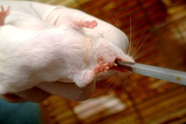
Fig- 2: Rosemary Extract administration via oral gavage